Contents include:
1. Structure of the human eye
2. Diseases of the eye
3. Preventions and treatments for these diseases
4. Fun and interesting animations and pictures, including optical illusions!
5. Discussions and controversial issues regarding diseases of the human eye
6. And many more links and resources for you to research on your own (:
Created on 27 February 2008 by 4 Biology students (:
& best viewed using Internet Explorer Version 7 and above or Mozilla Firefox.
We'll be blogging in the perspectives of different people:
Yee Herng as an Ophthalmologist (eye surgeon),
Xiu Li as an Eye Refraction Defect Specialist
Xiu Han as a Corneal Specialist,
and Wen Yi as a Student majoring in Biology.
Here is a type of test carried out to see if a person is colourblind.
What numbers can you see in the following images?
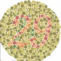
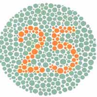
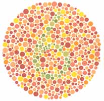
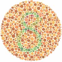
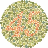
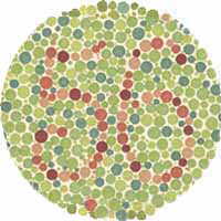
Answers: 29, 25, 6, 45, 8, 56
Did you see all of the numbers above? If you do, congratulations! Your vision is normal! If not, you should consult a doctor for a more detailed analysis.
Labels: ColourBlindness
0 Comments:
Fun Optical Illusions!
Primrose's field
This amazing illusion to the right was created by Akiyoshi Kitaoka. It already looks quite 'wavy', but try scrolling down slowly while concentrating on the image. You should see the picture 'waving' in an incredible way.

Wheel of Fun
Focus on the dot in the centre and then move your head towards and away from the monitor. You should see the circles rotate spookily! Sakura
Try scrolling down slowly while focusing on the middle of the picture. It looks like it's moving!
Flickering Fog
Based on an illusion byAkiyoshi Kitaoka. Stare intently at the red dot in the center of the left block for about a couple of minutes.Once you've done this, look over to the center of the right block. It should act weird - by fading out and 'flickering'. If you move your head closer and further from the monitor (while focusing on the center), you should see some interesting effects too.
Done By: Yee Herng
Labels: OpticalIllusions
2 Comments:
Cool illusions! :}
By  -xiuli-, at
March 9, 2008 at 4:53 PM
-xiuli-, at
March 9, 2008 at 4:53 PM
Yeah, cool illusions.. ^^
By \\wenyi//, at
March 10, 2008 at 12:03 AM
Types of colour blindness:
Red-green hereditary photoreceptor disorders [most common]
- Result from partial or complete loss of function in one or more of the different cone systems.
- The most frequent forms of human colour blindness result from problems with either the middle or long wavelength sensitive cone systems, and involve difficulties in discriminating reds, yellows and greens from one another. This is collectively referred to as "red-green colour blindness". However, this term is an over-simplification and is somewhat misleading.
- Other forms of colour blindness are much more rare. They include problems in differentiating blues from yellow, and the rarest forms of all, complete colour blindness or monochromacy, where one cannot distinguish any colour from grey, as in a black-and-white movie or photography.
Acquired colour blindness through damage to the eye, nerves or brain
- Unlike genetic disorders, acquired colour blindness may only occur in a portion of the visual field but maintain normal colour vision elsewhere
- Some forms are reversible
- Transient colour blindness, for migraine sufferers [very rare]
Classification of colour deficiencies
Colours of the rainbow as viewed by a person with no colour vision deficiencies

Colours of the rainbow as viewed by a person with protanopia

Colours of the rainbow as viewed by a person with deuteranopia

Colours of the rainbow as viewed by a person with tritanopia

Colour vision deficiencies can be classified:[as mentioned earlier].
- Acquired
- Inherited
Monochromacy:
- Also known as "total colour blindness".
- The lack of ability of distinguish colours
- Caused by cone defect or absence
- Occurs when two or all three of the cone pigments are missing and colour and lightness vision is reduced to one dimension.
Rod monochromacy:
- Rare, non-progressive inability to distinguish any colours as a result of absent or non-functioning retinal cones
- Associated with
- Light sensitivity [photophobia]
- Involuntary eye oscillations [nystagmus]
- Poor vision
Cone monochromacy:
- A rare, total colour blindness
- Accompanied by relatively normal vision, electroretinogram and electrooculogram
Dichromacy:
- A moderately severe colour vision defect
- One of the three basic colour mechanisms is absent or not functioning
- Hereditary
- Gender-linked - affects only males
- Occurs when one of the cone pigments is missing and colour is reduced to two dimensions
Protanopia:
- Severe type of colour vision deficiency caused by the complete absence of red retinal photoreceptors
- A form of dichromatism in which red appears dark
- Hereditary
- Gender-linked - occurs in 1% of all males
Deuteranopia:
- Colour vision deficiency in which the green retinal photoreceptors are absent, moderately affecting red-green hue discrimination
- A form of dichromatism in which there are only two types of cone pigments present
- Hereditary
- Gender-linked - present in 1% of all males
Tritanopia:
- Exceeding rare colour vision disturbance
- Only two cone pigments present
- A total absence of blue retinal receptors
- Anomalus trichromacy
- Common type of inherited colour vision deficiency
- Occurs when one of the three cone pigments is altered in its spectral sensitivity
- Results in an impairment, rather than a loss, of trichronmacy [normal three-dimensional colour vision]
Protanomaly:
- Mild colour vision defect
- An altered spectral sensitivity of red retinal receptors [closer to green receptor response] resulting in poor red-green hue discrimation
- Hereditary
- Gender-linked - present in 1% of all males
- Often passed from mother to child
Deuteranomaly:
- Caused by a similar shift in the green retinal receptors
- The most common type of colour vision deficiency
- Affects red-green hue discrimation in 5% of all males
- Hereditary
- Gender-linked
Tritanomaly:
- Rare
- Hereditary
- Colour vision deficiency affecting blue-yellow hue discrimation
Done by: Wen Yi
Labels: ColourBlindness
4 Comments:
Wow. I didn't know there were so many types of color deficiencies. How about those which people who are affected can only see in black and white? Is it the main type of color blindness?
By  Anonymous, at
March 9, 2008 at 4:28 PM
Anonymous, at
March 9, 2008 at 4:28 PM
Nope. Actually, people who can only see in black and white is the rarest form of colour blindness.
By \\wenyi//, at
March 9, 2008 at 11:44 PM
Good overview! I didn't know colour blindness can be passed on from mother to child.
A question for you: Can colour blindness be cured? Can the defect or absent cones be replaced?
By -xiuli-, at
March 10, 2008 at 10:50 AM
Generally, it can't be treated. But there are tinted contact lenses that can help the person suffering from colour blindness see better. A singular, red-tint contact lens can also be prescribed by the optometrists to wear in the dominant eye and can help the person to pass colour blindness tests for some occupations. Computer software has also been developed for people suffering from colour blindness to see better.
By \\wenyi//, at
March 10, 2008 at 7:49 PM
Introduction to colour blindness
What is it?
- The inability to spot the differences in some of the colours that can be spotted by other people.
Causes of colour blindness
- Often caused by genes
- But other possible causes are damage to the eye, nerve or brain.
Trivia facts about colour blindness
- Colour blindness is sometimes referred to as “Daltonism”, due to John Dalton’s work on his own colour blindness. However, this term is now used to describe a type of colour blindness known as “deuteranopia”.
- Colour blindness is classified as a disability.
- Colour blind people can have an advantage over people with normal colour vision in certain situations.
- Some studies show that colour blind individuals are better at seeing through certain camouflages. *Monochromats may have a minor advantage in dark vision, but only in the first five and a half minutes of dark adaptation.
The following is an 1895 illustration of normal vision and various kinds of color blindness.
 *Refer here for more information on monochromats.
Background information
Color Blindness affects both men and women. The normal human retina contains two kinds of light cells: the rod cells (active in low light) and the cone cells (active in normal daylight).
Normally, there are three kinds of cones, each containing a different pigment. The cones are activated when the pigments absorb light. The absorption spectra of the cones differ; one is maximally sensitive to short wavelengths, one to medium wavelengths, and the third to long wavelengths (their peak sensitivities are in the blue, yellowish-green, and yellow regions of the spectrum, respectively).
The absorption spectra of all three systems cover much of the visible spectrum, so it is not entirely accurate to refer to them as "blue", "green" and "red" receptors, especially because the "red" receptor actually has its peak sensitivity in the yellow.
The sensitivity of normal color vision actually depends on the overlap between the absorption spectra of the three systems: different colors are recognized when the different types of cone are stimulated to different extents. Red light, for example, stimulates the long wavelength cones much more than either of the others, and reducing wavelength causes the other two cone systems to be increasingly stimulated, causing a gradual change in hue.
Done by: Wen Yi
Labels: ColourBlindness
6 Comments:
You said that "Color Blindness affects both men and women."
However, colour blindness affects more women or men? If I'm not wrong, colour blindness happens to one of the genders more than the other.
By -xiuli-, at
March 9, 2008 at 5:00 PM
Maybe, you want to re-upload your pic. It's kind of blur. Take from
http://en.wikipedia.org/wiki/Image: US_Flag_color_blind.png
This one is enlarged size so it will be clearer, I think. >:D
By -xiuli-, at
March 9, 2008 at 5:03 PM
In some forms of colour blindness, more men are affected than women. Refer to the latest post for details.
By \\wenyi//, at
March 10, 2008 at 12:35 AM
This comment has been removed by the author.
By -xiuli-, at
March 10, 2008 at 10:42 AM
Okay. Just curious. Does the eyes of a colour blindness sufferer look different? Or, the difference can't actually be noticed?
By -xiuli-, at
March 10, 2008 at 10:54 AM
The eyes of a colour blindness will not look any different from that of a normal person's. So unless the person reveals that he/she is colourblind, we cannot tell that the person is colourblind.
By \\wenyi//, at
March 10, 2008 at 8:04 PM
For more information, click here for a clear overview of myopia and hyperopia.
The site includes surgical treatments such as LASIK and PRK and is in swf format (a flash animation).
0 Comments:
CORNEAL TRANSPLANTATION
I will be talking about corneal transplantation. Corneal transplantation is also known as corneal grafting, which is a surgical procedure where a damaged or diseased cornea is replaced by donated corneal tissue which has been removed from a recently deceased individual having no known diseases which might affect the viability of the donated tissue.
The first cornea transplant was performed in 1905, by Eduard Zirm, making it one of the first types of transplant surgery successfully performed.
Corneal transplantations are done to improve visual acuity by replacing the opaque host tissue by clear healthy donor tissue; to preserve corneal anatomy and integrity in patients with stromal thinning and descemetoceles, or to reconstruct the anatomy of the eye; to remove inflamed corneal tissue unresponsive to antibiotics or anti-virals treatments and to improve the appearance of patients with corneal scars that have given a whitish or opaque hue to the cornea.
On the day of the surgery, the patient is given a brief physical examination by the surgical team and is taken to the operating room and given anesthesia.
With anesthesia induced, the surgical team prepares the eye to be operated on. An eyelid speculum is placed to keep the lids open, and some lubrication is placed on the eye to prevent drying. A metal ring is then stitched to the sclera, which will provide a base for a trephine.
A trephine is then placed over the cornea and is used by the surgeon to cut the host cornea. The trephine is then removed and the surgeon cuts a circular graft from the donor cornea. Once this is done, the surgeon returns to the patient's eye and removes the host cornea.
The donor cornea is then brought into the surgical field and maneuvered into place with forceps.
Once in place, the surgeon will fasten the cornea to the eye. The surgeon finishes up by reforming the anterior chamber with a sterile solution injected by a canula, then testing that it's watertight by placing a dye on the wound exterior.
With the metal ring removed and antibiotic eyedrops placed, the eye is patched, and the patient is taken to a recovery area while the effects of the anesthesia wear off. The patient typically goes home following this and sees the doctor the following day for the first post operative appointment.
Labels: CorneaTransplant
1 Comments:
You said "..where a damaged or diseased cornea is replaced by donated corneal tissue.."
How did the cornea become diseased or damaged in the first place actually?
By -xiuli-, at
March 9, 2008 at 4:51 PM
Hyperopia
swf file taken from allaboutvision.com & requires flash player to view
-XiuLi
Labels: Hyperopia
0 Comments:
Signs and symptoms of hyperopia
• general eye discomfort or a headache after prolonged interval of doing close tasks, such as reading, writing or drawing
• eyestrain, including burning eyes, aching in or around the eyes, and, rarely, a headache
• squinting of eyes to see near objects clearer
Treatment of hyperopia
People with hyperopia often squint when working with objects up close. This allows them to see better as a different amount of light is refracted. Like treatments for myopia, treatments for hyperopia are designed to refract a correct amount of light into the eyes through either corrective lenses or reshaping of the cornea through surgical and non-surgical methods.
Like myopia, spectacles, contact lenses, and refractive surgery are the primary options to correct the refraction error of hyperopia.
Similarly, eyeglasses and contacts are usually the first treatment people use for hyperopia.
However, the inconveniences of using spectacles and contacts have lead to the search for other treatments.
Depending on the degree of the patient's myopia and his/her age, other techniques may be used. Laser assisted in situ keratomileusis, or LASIK Eye Surgery, is the most popular surgery used to treat hyperopia.
• In this procedure the shape of the cornea is measured and changed to fix the degree of myopia using a laser.
An alternative to LASIK, Conductive Keratoplasty, CK for short, uses the same concept of reshaping the cornea but uses different procedures and caters to patients who are over 40 and have hyperopia in the range from +0.75 to +3.00 diopters.
For patients who have extreme myopia or hyperopia, there's a new treatment, Phakic Intraocular Lenses that can be used to correct high degrees of hyperopia and is also a promising option for patients with hyperopia who also suffer from presbyopia.
• uses an implantation of lens much like a contact lens inside the eye to allow better light refraction
• correct the refractive error without removing the natural lens
-XiuLi
Labels: Hyperopia
0 Comments:
Trachoma
Trachoma (Ancient Greek: "rough eye") is an infectious eye disease, unlike cataracts which are mainly caused by exposure to radiation.
Globally, 84 million people suffer from active infection and nearly 8 million people are visually impaired as a result of this disease. However, few people have heard of trachoma. It is often overlooked as a priority for public-health intervention because it is not fatal. In rural communities, people live in overcrowded conditions with limited access to water and health care. In some communities, the disease is so common that blindness from trachoma is simply accepted as a fact of life. Yet trachoma is treatable, and the suffering it causes is avoidable.
Those who are infected by trachoma do not instantly go blind. The disease manifests gradually. Children may not notice its effects until adulthood, when scarring from repeated infections causes the eyelashes to turn inward and scratch the cornea, leading slowly to complete blindness.

Causes
Trachoma is caused by the bacterium Chlamydia trachomatis and it is spread by direct contact with eye, nose, and throat secretions from affected individuals, or contact with inanimate objects such as towels or washcloths that have had similar contact with these secretions. The bacteria can be spread easily on an infected person's hands or clothing, or may be carried by pests that were in contact with the discharge of an infected person. Children are most susceptible to infection, but the effects are often not felt until adulthood.
Because trachoma is transmitted through close personal contact, it tends to occur in clusters, often infecting entire families and communities. Women are up to three times more likely than men to be blinded by the disease.
Symptoms
The bacteria Chlamydia trachomatis has an incubation period of 5 to 12 days, after which the affected individual experiences symptoms of conjunctivitis, or irritation similar to "red eye".
Further symptoms include:
- Eye discharge
- Swollen eyelids
- Trichiasis (turned-in eyelashes)
- Swelling of lymph nodes in front of the ears
- Corneal scarring
- Further ear, nose and throat complications.

Although trachoma was eliminated from much of the developed world in the last century, this disease persists in many communities without adequate access to water and sanitation. Trachoma keeps families shackled within a cycle of poverty, as the disease and its long-term effects are passed from one generation to the next.
The World Health Organization (WHO) has set a goal of eliminating blinding trachoma as a public health concern by 2020. National governments in collaboration with numerous non-profit organizations implement trachoma control programs using the WHO-recommended SAFE strategy, which includes:
- Surgery to correct advanced stages of the disease
- Antibiotics to treat active infection, using Zithromax
- Facial cleanliness to reduce disease transmission
- Environmental change to increase access to clean water and improved sanitation.
Effects
If not treated properly with oral antibiotics, the symptoms may escalate and cause blindness, which is the result of ulceration and consequent scarring of the cornea. Surgery may also be necessary to fix eyelid deformities.
 Nerve signals travel from each eye along the optic nerve and other nerve fibers (called the visual pathway) to the back of the brain, where vision is sensed and interpreted. at the optic chiasm (in front of the pituitary gland), the optic nerve from each eye divides, and half of the nerve fibers cross to the other side to the back of the brain. Thus, the right side of the brain receives information through both optic nerves for the left field of vision, and vice versa. The middle of these fields of vision overlaps. It is seen by both eyes (called binocular vision).
An object is seen from slightly different angles by each eye so the information the brain receives from each eye is different, although it overlaps. The brain integrates the information to produce a complete picture.
Done By: Yee Herng
Labels: Blindness
0 Comments:
The opposite defect of myopia is hyperopia.
What is hyperopia?
• also known as hypermetropia or as farsightedness or longsightedness
• a common vision condition in which you can see objects in the distance clearly, but objects nearby may be blurry
• with mild farsightedness, one may see clearly objects that are closer
• with severe farsightedness, one can see clearly only objects a great distance away
• in extreme cases, one can’t even focus on objects at any distance
Causes of hyperopia?
• caused when the cornea is too flat (curved too little) or when the eye is too short
How or why hyperopia happens?
simply put, the light entering the eyes is not focused correctly, focused behind the retina rather than directly on the retina
• refractive defect of the eye causes collimated light to produce image focus behind the retina
Note: Hyperopia is often confused with presbyopia. People suffering from presbyopia will also experience blurry near vision and good far vision. However, these are two different eye conditions brought about by different causes.
___________________________________________________________________
Presbyopia is an eye condition that will happen to some degree to everyone some time in life. It in inevitable and there is no cure for presbyopia although there are a lot of procedures to help correct presbyopia to allow improved near vision.
It occurs because of a reduced accommodative amplitude which is brought about by natural aging changes with the crystalline lens. As we age, the lens of the eyes becomes rigid, loses its flexibility, and thus its ability to focus as well, causing presbyopia.
-XiuLi
Labels: Hyperopia
2 Comments:
"With mild farsightedness, one may see clearly objects that are closer." Do you mean that mild farsightedness (hyperopia) is similar to myopia, or that a person who has mild hyperopia can see close objects clearer than those with severe hyperopia? Can you elaborate more on this point? thanks (:
By yh., at
March 9, 2008 at 5:59 PM
Nope. Sorry, I didn't make it clearer.
I meant to say that as compared to a more severe case of farsightedness, one with mild hyperopia will be able to see clear images from a closer distance than that of the person suffering from severe hyperopia.
Hyperopia is NOT like myopia at all. It is literally its opposite.
When suffering from hyperopia, near objects appear blurred while when suffering from myopia, far objects appear blurred. Hence, the names given.
Myopia, which is nearsightedness, is when near objects appear clear and hyperopia, which is farsightedness, is when far objects appear clear to the eye.
By -xiuli-, at
March 9, 2008 at 6:17 PM
Myopia
swf file taken from allaboutvision.com & requires flash player to view
-XiuLi
Labels: Myopia
0 Comments:
Myopia -introduction
" Myopia is becoming very common. In Singapore, the rate of myopia amongst school kids has increased from about 25% just 30 years ago to about 75%. "
What is myopia?
• many different types i.e. pathological myopia, secondary myopia, pseudomyopia, nocturnal myopia and the most common simple myopia
___________________________________________________________________
Simple myopia
• also called nearsightedness or short sightedness
• a vision condition in which near objects are seen clearly but distant objects do not come into proper focus, appearing blurred
Causes of simple myopia?
• caused by a natural change in shape of the eyeball that makes it egg-shaped rather than round
• if the eyeball is too long or the cornea has too much curvature (too steep)
How or why simple myopia happens?
simply put, the light entering the eyes is not focused correctly, focused in front of the retina rather than directly on the retina
• light focused on the vitreous humour inside the eye rather than on the retina at the back of the eye
• refractive defect of the eye causes collimated light to produce image focus in front of the retina when accommodation is relaxed
___________________________________________________________________
Nocturnal myopia
• also known as twilight myopia or night myopia
• a condition in which the eye has a great difficulty seeing in low illumination areas although daytime vision is normal
___________________________________________________________________
Pseudomyopia
• the blurring of distance vision brought along by the spasm of the ciliary muscle
• common in young adults
Causes of pseudomyopia?
• differs from simplel myopia in that the focusing of light in front of the retina is due to transient spasm of the ciliary muscle which causes an increase in the refractive power of the eye
• classically occurs after change in visual requirements, such as students preparing for an exam, or a change in occupation
• can only be caused by diseases such as uncontrolled diabetes, Myasthenia gravis and nervous system disorders
-XiuLi
Labels: Myopia
2 Comments:
Hmm. Are you trying to say that Pseudomyopia is the rarest form of myopia, as it can only be caused by certain types of diseases such as untreated diabetes, and can only form after a changed occupation or lifestyle?
Anyway, great piece of information i learned after viewing your post. :D
By yh., at
March 9, 2008 at 4:40 PM
It is very common in young adults as written in the post as it can be caused by a lot of stress, especially from schoolwork.
By -xiuli-, at
March 9, 2008 at 4:44 PM
A cataract is an opacity that develops in the crystalline lens of the eye or in its envelope. Early in the development of age-related cataract, the power of the crystalline lens may be increased, causing near-sightedness (myopia), and the gradual opacification of the lens may reduce the perception of blue colours. Cataracts progress slowly to cause vision loss and are potentially blinding if untreated.
Cataract is derived from the Latin cataracta meaning "waterfall". Cataracts are the leading cause of blindness in the world. In the United States, age-related lenticular changes have been reported in 42% of those between the ages of 52 to 64, 60% of those between the ages 65 and 74, and 91% of those between the ages of 75 and 85.
Causes
Cataracts may be partial or complete, stationary or progressive, and hard or soft. Cataracts are also classified by their location, e.g. posterior and anterior [common (senile) cataract is related to aging].
1. Secondary cataract. Cataracts can form after surgery for other eye problems, such as glaucoma. Cataracts also can develop in people who have other health problems, such as diabetes. Cataracts are sometimes linked to steroid use.
2. Traumatic cataract. Cataracts can develop after an eye injury, sometimes years later.
3. Congenital cataract. Some babies are born with cataracts or develop them in childhood, often in both eyes. These cataracts may be so small that they do not affect vision. If they do, the lenses may need to be removed.
4. Radiation cataract. Cataracts can develop after exposure to some types of radiation.
How Cataracts Affect Vision
 On the left, a normal lens receives light and focuses it on the retina. On the right, a cataract blocks some light from reaching the lens and distorts the light being focused on the retina.
 How a person with cataract sees the world
Symptoms
The following symptoms can also be a sign of other eye problems such as myopia. If you have any of these symptoms, be sure to check with your eye care professional.
- Cloudy or blurry vision.
- Colors seem faded.
- Glare. Headlights, lamps, or sunlight may appear too bright. A halo may appear around lights.
- Poor night vision.
- Double vision or multiple images in one eye. (This symptom may clear as the cataract gets larger.)
- Frequent prescription changes in your eyeglasses or contact lenses.
Prevention
Although cataracts have no scientifically proven prevention, it is sometimes said that wearing ultraviolet-protecting sunglasses may slow the development of cataracts. Regular intake of antioxidants (such as vitamin A, C and E) is theoretically helpful, but taking them as a supplement has been shown to have no benefit.
Treatment
The most effective and common treatment is to surgically remove the cloudy lens. There are two types of surgery that can be used to remove cataracts: extracapsular cataract extraction (ECCE) and intracapsular cataract extraction (ICCE).
Extra-capsular (ECCE) surgery consists of removing the lens but leaving the majority of the lens capsule intact. High frequency sound waves are sometimes used to break up the lens before extraction.
Intra-capsular (ICCE) surgery removes the entire lens of the eye, including the lens capsule, but is rarely performed in modern practice. In either of the surgeries, the cataractous lens is removed and replaced with a plastic lens which stays in the eye permanently.
The internal structure of the eye. Click 'Introduction' on the navigations to find out more about our eye! (:

Done By: Yee Herng
Labels: Blindness
2 Comments:
Can cataracts spread from one eye to another?
I mean, if only your right eye has cataracts, will your left eye also suffer from cataracts because of infection from your right eye?
By -xiuli-, at
March 9, 2008 at 4:46 PM
Glad you brought up that point.
No, a cataract can occur in either or both eyes, but it cannot spread from one eye to the other.
It is rather difficult for inter-eye spreading of cataract because cataracts are mainly caused by radiation or protein clumping. (refer to 'causes' in the post)
By yh., at
March 9, 2008 at 5:38 PM
Signs and symptoms of myopia
• headaches caused by eyestrains
• squinting of eyes to see far objects clearer
• persistent squinting
• sitting only short distances from television, movie screen or whiteboard
• holding of books very closely while reading
• unawareness of distant objects
Treatment of myopia
People with myopia usually are able to see a little better by squinting. This is caused by the different amount of light refracted into the eyes. All treatments for myopia have this same goal. Spectables, contacts and different surgeries all have the goal of correcting this refraction error.
Spectacles, contact lenses, and refractive surgery are the primary options to correct the refraction error of myopia.
Eyeglasses and contacts are usually the first treatment people use for myopia.
However, the inconveniences of using spectacles and contacts have lead to the search for other treatments.
Depending on the degree of the patient's myopia, other techniques may be used. Laser assisted in situ keratomileusis, or LASIK Eye Surgery, is the most popular surgery used to treat myopia.
• In this procedure the shape of the cornea is measured and changed to fix the degree of myopia using a laser.
An alternative to LASIK, Photorefractive Keratectomy, PRK for short, uses the same concept of reshaping the cornea but uses different procedures.
For non-surgical treatments (except glasses and contacts), there are Orthokeratology and Corneal Ring Segments.
People with lower degrees of myopia are better suited for these techniques.
• Orthokeratology uses RGP contact lenses to gradually and temporarily reshape the curvature of the cornea.
• Corneal ring segments are small polymer plastic materials that are implanted into the cornea to reshape it for corrected vision.
Labels: Myopia
0 Comments:
Blindness - Loss of vision
Definition
Blindness is the condition of a lack of vision or the inability to see. It may also refer to a loss of vision that cannot be corrected with glasses or contact lenses.
Partial blindness means you have very limited vision. Complete blindness means you cannot see anything and do not see light. People with vision worse than 20/200 are considered legally blind in the United States.
Causes
Blindness has many causes. The leading causes of chronic blindness include cataract, glaucoma, age-related macular degeneration, corneal opacities, diabetic retinopathy, trachoma, and eye conditions in children (e.g. caused by vitamin A deficiency).
Age-related blindness is increasing throughout the world, as is blindness due to uncontrolled diabetes. On the other hand, blindness caused by infection is decreasing, as a result of public health action. Three-quarters of all blindness can be prevented or treated.
Most visual impairment is caused by disease and malnutrition. According to WHO estimates in 2002, the most common causes of blindness around the world are: cataracts (47.8%), glaucoma (12.3%), uveitis - inflammation involving the interior of the eye (10.2%), age-related macular degeneration (AMD) (8.7%), corneal opacity (5.1%), diabetic retinopathy - damage to the retina (4.8%), and trachoma - infectious eye disease (3.6%).
Other causes include:
- Blocked blood vessels
- Complications of premature birth (retrolental fibroplasia - abnormal blood vessel development in the retina in a premature infant)
- Complications of eye surgery
- Lazy eye
- Optic neuritis (inflammation of the optic nerve)
- Stroke
- Tay-Sachs disease (deadly disease passed down through families that causes damage to the nervous system)
- Retinitis pigmentosa (an eye disease which causes damage to the retina. Causes problems with night vision and peripheral vision.)
- Retinoblastoma (cancer of the retina, affects children under age of 6)
- Lead poisoning
- Optic glioma (tumors that grow in various parts of the brain)
I will focus on two different causes of blindness - Cataracts and Trachoma in the later posts.

 The world looks different to an insect because of the compound nature of its eyes and also because they are more sensitive to different wavelengths of light than our eyes are. For example some insects can see UV rays. Features of plants that are invisible to our eyes are apparent to insects because the patterns of the features are visible in the UV rays.
Done by: Yee Herng
Labels: Blindness
5 Comments:
What is glaucoma?
It is related to cataracts right?
By -xiuli-, at
March 9, 2008 at 4:48 PM
Glaucoma is optic nerve damage, often associated with increased eye pressure, that leads to progressive, irreversible loss of vision.
In glaucoma, the canals through which the fluid drains become clogged, blocked, or covered. Fluid cannot leave the eye even though new fluid is being produced in the posterior chamber. Because there is nowhere in the eye for the fluid to go, pressure in the eye increases. When the pressure becomes higher than the optic nerve can tolerate, glaucoma results.
It usually has nothing to do with cataracts, and is just another cause of blindness.
For more information, you can check out 'glaucoma' under 'resources' in the 'credits' section.
By yh., at
March 9, 2008 at 5:06 PM
Okay. :) Now, I understand.
But, secondary cataract can be caused by glaucoma right?
By -xiuli-, at
March 9, 2008 at 5:22 PM
In the 'Cataracts' post, I mentioned that cataracts can form after surgery for other eye problems, such as glaucoma. But cataracts are usually not caused by glaucoma itself, rather, after the surgery for eye problems (infection). So the answer is no, as cataracts are not caused by glaucoma itself.
By yh., at
March 9, 2008 at 5:43 PM
O, I see. You cleared my queries!
By -xiuli-, at
March 9, 2008 at 5:48 PM

This is the vertical cut of the eye, where you can see EVERYTHING inside the eye! =P
The outer layer: the Sclera and Cornea
The sclera is the tough, fibrous outer layer of the eyeball that forms the whites of your eyes. The front of the sclera is covered by the conjunctiva — a thin, transparent membrane that’s involved in protecting your eyes. The conjunctiva also lines the insides of the eyelids. The cornea is a transparent dome-shaped structure at the front of the eye allowing light to enter the eye, and helps focus and direct light onto the retina together with the lens.
The middle layer: the uveal tract (iris, ciliary body and choroid) and lens
The iris is the coloured part of your eye that controls the size of the pupil — the black area in the centre of the iris. When you are in bright light, the iris reduces the pupil size to restrict the amount of light entering the eye; when in dim light or darkness, the iris opens up the pupil to allow more light in. the iris is between the posterior and anterior chamber, which contains aqueous humour which is a watery liquid formed by the ciliary body constantly. It keeps the lens and cornea nourished.
The lens is a clear, flexible structure that changes shape so that you can focus on objects at varying distances. It is connected to the ciliary body by suspensory ligaments, and changes it's length and thickness according to the relaxation and contraction of ciliary muscles to view objects at different distances.
The vitreous humour is a jelly-like substance that fills the back portion of the eye behind the lens. It helps the eye to keep its shape, and transmits light to the back of the eye.
The choroid is a membrane between the sclera and the retina that lines the back of the eye. It contains many blood vessels that supply oxygen and nutrients to the retina, and is highly pigmented to help absorb light and prevent scattering.
The inner layer: the retina
The retina lines the inside of the back part of the eye, and is light-sensitive part. It contains millions of cells known as photoreceptors, and each is linked to a nerve fibre. You have a blind spot, where all of these nerve fibres converge to form the optic nerve. Objects that fall on the blind spot of one eye are seen by the other eye.
Once an image is detected by the photoreceptors, this information is converted into nerve impulses that are sent to the brain via the optic nerve.
The macula is a small area of the retina that contains a high concentration of photoreceptors, and the middle part of the macula — the fovea — is the most sensitive area, providing the sharpest vision.
So after my long explanation, do you know more about our eyes? =)
-xh
These are some questions you may have on the eye. If you have any questions, feel free to drop a mail and i will try to answer! smilez!
Q: How does normal vision develop?
A:
Newborn infants are able to see, but as they use their eyes during the first months of life, vision improves.
During early childhood years, the visual system changes quickly and vision continues to develop. If a child cannot use his or her eyes normally, vision does not develop properly and may even decrease. After the first nine years of life, the visual system is usually fully developed and usually cannot be changed.
The development of equal vision in both eyes is necessary for normal vision. Many occupations are not open to people who have good vision in one eye only. If the vision in one eye should be lost later in life from an accident or illness, it is essential that the other eye have normal vision. Without normal vision in at least one eye, a person is visually impaired.
Q: Why does eyelids twitch sometimes?
A: Mild twitching of the eyelid is common. Though annoying, they are almost always temporary and completely harmless. When your eye is twitching, it is not visible to anyone else. Lack of sleep, too much caffeine or increased stress seem to be root causes of twitching. Often, gently massaging your eye will relieve the symptoms. Usually, the twitch will disappear after catching up on your sleep...so go and sleep if it's way past your bedtime! =)
Q: What is legal blindness?
A: You are legally blind when the best corrected central acuity is less than 20/200 (perfect visual acuity is 20/20) in your better eye, or your side vision is narrowed to 20 degrees or less in your better eye. Even if you are legally blind, you may still have some useful vision. If you are legally blind, you may qualify for certain government benefits.
Q: What is visual impairment?
A: If neither of your eyes can see better than 20/60 without improvement from glasses or contacts, you may be defined as visually impaired. In addition, poor night vision, limited side vision, double vision and loss of vision in one eye may also determine visual impairment.
Q: What is low vision?
A: Low vision is a term describing a level of vision below normal (20/70 or worse) that cannot be corrected with conventional glasses. Low vision is not the same as blindness. People with low vision can use their sight. However, low vision may interfere with the performance of daily activities, such as reading or driving.
Q: Will working at a computer screen hurt my eyes?
A: No, there is no evidence that working at a computer damages the eyes. However, long hours of work can be fatiguing to the eyes, neck and back. Monitor glare from various light sources can also be a problem. It is often helpful to take periodic breaks, looking off in the distance and adjusting your work station (angle of the monitor, height of the chair, changing the lighting).
Q: How does the eye work?
A: The front parts of the eye (the cornea, pupil and lens) are clear and allow light to pass through. The light also passes through the large space in the center of the eye called the vitreous cavity. The vitreous cavity is filled with a clear, jelly-like substance called vitreous humour. The light is focused by the cornea and the lens onto a thin layer of tissue called the retina, which covers the back inside wall of the eye. The retina is like the film in a camera. It is the seeing tissue of the eye. When the focused light hits the retina, a picture is taken. Messages about this picture are sent to the brain through the optic nerve. This is how we see.
-xh
Links to learn more, with more in-dept analysis.
Glaucoma
Cataracts
Sight
Credits to these websites we used for references
information:
Bee/Insect View
Optical Illusions
Cataracts
Myopia -1
Myopia -2
Hyperopia
Presbyopia
Colour blindness 1
Colour blindness 2
uploading:
background image
swf files
layout: detonatedlove edited by:xiuli
background image: collated by yeeherng
inspiration: wordpress